Kamis, 07 Januari 2010

Amino Acid Metabolism

This page describes the amino acids that are important in human nutrition. It covers the digestion and absorbtion of proteins in the gut, some of the amino acid degradation pathways in the tissues, and the urea cycle. There are separate pages dealing with nucleotide and 'one carbon' metabolism and with porphyrin metabolism. Other aspects of amino acid biochemistry are covered in the section on the integration of metabolism. Click here to skip over the detailed contents section.

Protein turnover

The recommended minimal protein intake required to achieve nitrogen balance in healthy adults is about 50g per day, although in developed countries many people may eat double this amount. This compares with an average daily protein turnover of about 250g per day. Human proteins have very different lifetimes. Total body protein is about 11kg, but about 25% of this is collagen, which is metabolically inert. A typical muscle protein might survive for three weeks, but many liver enzymes turn over in a couple of days. Some regulatory enzymes have half-lives measured in hours or minutes. The majority of the amino acids released during protein degradation are promptly re-incorporated into fresh proteins. Net protein synthesis accounts for less than one third of the dietary amino acid intake, even in rapidly growing children consuming a minimal diet. Most of the ingested protein is ultimately oxidised to provide energy, and the surplus nitrogen is excreted, a little as ammonia but mostly as urea.

Soluble intracellular proteins are tagged for destruction by attaching ubiquitin, a low molecular weight protein marker. They are then degraded in proteasomes to short peptides. A very few of these are displayed on the cell surface by the MHC [major histocompatibility] complex as part of the immune system, but most of them are further metabolised to free amino acids. Some proteins are degraded by an alternative system within the lysosomes.

Dietary proteins are initially denatured by the stomach acid, in conjunction with limited proteolysis by pepsin. In young mammals gastric rennin [do not confuse with renin!] partially hydrolyses and precipitates milk casein and increases gastric residence time. Gastric acid also kills most ingested bacteria, rendering the upper part of the gut almost sterile.

Protein digestion is largely completed in the small intestine at a slighlty alkaline pH. The pancreatic proteases trypsin, chymotrypsin and elastase divide the proteins into short peptides. These are attacked from both ends by aminopeptidase and carboxypeptidase, and the fragments are finished off by dipeptidases secreted from the gut wall.

Amino acid uptake from the gut lumen into enterocytes is driven by the sodium gradient. There is a relatively high sodium concentration in the gut (regardless of dietary intake, as a result of the pancreatic secretion of sodium bicarbonate) and a low concentration in the enterocytes, as a result of the sodium pump in the basolateral membrane. A multiplicity of sodium-linked amino acid carriers operate within the intestinal brush border, balanced by sodium-independent export carriers on the serosal surface (i.e. the opposite side) of the cells.

Central role of glutamate

Four of the amino acids: glutamate, aspartate, alanine and glutamine are present in cells at much higher concentrations than the other 16. All four have major metabolic functions in addition to their roles in proteins, but glutamate occupies the prime position.

Glutamate and aspartate function as excitatory neurotransmitters in the central nervous system, and glutamate is partly responsible for the flavour of food. (It is the mono sodium glutamate listed on processed food labels.) However, glutamate also occupies a special position in amino acid breakdown, and most of the nitrogen from dietary protein is ultimately excreted from the body via the glutamate pool.

Glutamate is special because it is chemically related to 2-oxoglutarate (= alpha keto glutatarate) which is a key intermediate in the citric acid (Krebs) cycle. Glutamate can be reversibly converted into oxoglutarate by transaminases or by glutamate dehydrogenase. In addition, glutamate can be reversibly converted into glutamine, an important nitrogen carrier, and the most common free amino acid in human blood plasma.

Transamination reactions

Most common amino acids can be converted into the corresponding keto acid by transamination. This reaction swops the amino group from one amino acid to a different keto acid, thereby generating a new pairing of amino acid and keto acid. There is no overall loss or gain of nitrogen from the system - it is simply a question of "robbing Peter to pay Paul".

Transamination reactions are readily reversible, and the equilibrium constant is close to 1. One of the two pairs is almost invariably glutamate and its corresponding keto acid oxoglutarate, although there are a few exceptions to this rule. All transaminases require pyridoxal phosphate (derived from vitamin b6) as a cofactor.

The substrates bind to the active centre one at a time, and the function of the pyridoxal phosphate is to act as a temporary store of amino groups until the next substrate comes along. In the process the pyridoxal phosphate is converted into pyridoxamine phosphate, and then back again. Enzymologists call this a "ping pong" mechanism, and it leads to a characteristic pattern in the reaction kinetics.

The condensation between the alpha amino group and the aromatic aldehyde to form a "Schiff base" makes the alpha carbon atom chemically reactive, so the isomerisation of the Schiff base takes place very easily. In practice the pyridoxal form of the coenzyme condenses with the epsilon amino group of a lysine residue in the enzyme protein when no amino acid is bound, and the free aldehyde form of the coenzyme has only a transitory existence. Many of the enyzmes that metabolise amino acids require pyridoxal phosphate as a cofactor. Unexpectedly, this compound also serves in a different manner in the active centre of glycogen phosphorylase.

Glutamate:oxaloacetate transaminase [GOT]

This enzyme is also known as aspartate aminotransferase and is one of the most active enzymes in the cell. It exists in mitochondrial and cytosolic variants, and the detailed iso-enzyme pattern is tissue-specific. It escapes in large amounts from dead or dying tissues and enters the bloodstream, so GOT is often measured in blood samples for medical diagnostic purposes.

The metabolic importance of this enzyme is that it brings about a free exchange of amino groups between glutamate (which is the most common amino acid) and aspartate which is a second major amino acid pool. Glutamate and aspartate are each required for separate but essential steps in the urea cycle, which is responsible for ammonia detoxication and nitrogen excretion. The free movement of nitrogen between the glutamate and aspartate pools is an important balancing process that is vital for normal cellular metabolism.

This reaction is close to equilibrium in both the cytosol and the mitochondrial compartments. It forms an integral part of the malate - asparate shuttle which is effectively responsible for the "transport" of NADH across the inner mitochondrial membrane.

Glutamate:pyruvate transaminase [GPT]

This very active enzyme is also known as alanine aminotransferase and exists in mitochondrial and cytosolic variants. The detailed iso-enzyme pattern is tissue-specific. It escapes in large amounts from dead or dying tissues and GPT may be measured in blood samples for medical diagnostic purposes.

Alanine is the principal amino acid released from muscle tissue during starvation. It is an important substrate for hepatic gluconeogenesis, and alanine transamination is required for the proper maintenance of fasting blood glucose concentrations.

Glutamate dehydrogenase [GluDH]

This enzyme is the first committed step on the final common pathway for mammalian nitrogen excretion, leading eventually to urea. A few of the amino acids have specific deamination pathways, but about 75% of ingested protein nitrogen follows the glutamate route.

Glutamate dehydrogenase in mammals is almost entirely confined to the liver mitochondrial matrix space, where it accounts for a significant proportion of the total protein. In contrast to the transamination reactions which merely swop amino groups from one compound to another, GluDH catalyses a net loss of nitrogen from the amino acid pool. The process is therefore termed "oxidative deamination". It is the only common dehydrogenase which is non-specific for NAD or NADP, and this may be important for its overall regulation.

NADH / NAD and NADPH / NADP have the same standard redox potential of -420mV when the oxidised and reduced forms are present in equal concentrations. In practice these coenzymes have different effective redox potentials and perform specialised functions within cells. The NADPH / NADP pool operates almost entirely in the reduced form, but the NADH / NAD pool is rarely more than 30% reduced. In general NADPH is used to drive reductive biosynthetic reactions, whereas NAD is the coenzyme for the oxidative energy-yielding pathways.

The dual coenzyme specificity is a potential source of difficulty for the cell, since in theory this readily reversible enzyme could catalyse a futile cycle, proceeding first in the oxidative direction with NAD, followed by a reductive step using NADPH. The effect would be to "short circuit" the two coenzyme pools, which normally require considerable investment in substrates and cellular equipment to keep them separate. If this futile cycle happens to any significant extent then it must be an advantage for the cell, because it has persisted unchanged for 2,000,000,000 years of evolutionary development.

The most likely explanation at present is that this futile cycle takes place, but for various reasons it does not place an excessive burden on its owner. The Km of GluDH for ammonia is quite high, and the free ammonia concentration is kept very low by the next enzyme in the pathway, carbamyl phosphate synthetase. This will severely reduce the rate of the synthetic reaction, and allow the enzyme to catalyse a net glutamate oxidation at a slow controlled rate that provides the maximum opportunity for regulatory interference.

Regulation is plainly critical at this point, since GluDH and carbamyl phosphate synthetase jointly control the overall rate of nitrogen excretion and determine whether a particular individual will be in positive, neutral or negative nitrogen balance. Control of unwanted nitrogen losses remains an important unsolved problem after major surgery, burns or other serious traumatic injuries.

The enzyme is indeed modulated by adenine and guanine nucleotides, although it is difficult to make much sense of the observed effects. GluDH has all the hallmarks of a large multimeric allosteric enzyme, although the true nature of the regulation

Remains to be identified. The situation is in some ways similar to the parallel NAD and NADP linked oxidation pathways for malate and isocitrate, although the competing reactions for these substrates are separately regulated and catalysed by different proteins.
The liver GluDH gene is located on chromosome 10. OMIM link Surp

risingly few genetic defects have been reported, possibly reflecting the catastrophic nature of the resulting fault. There are reports of an additional GluDH gene located on the X chromosome OMIM link which is expressed in testis and neural tissues. This seems to be a processed pseudogene that is still functional.

Trans-deamination

Most transaminases share a common substrate and product (glutamate and oxoglutarate) with glutamate dehydrogenase, and this permits a combined nitrogen excretion pathway for individual amino acids that is commonly described as "trans-deamination".

This process underlines the central role of glutamate in the overall control of nitrogen metabolism.

Urea cycle

Ammonium ions are in equilibrium with about 1% free ammonia at physiological pH. Ammonium salts are toxic compounds, causing vomiting, convulsions and ultimately coma and death when the blood concentration exceeds approximately 0.25mM. It is not entirely clear why this should be so: it may be that ammonium ions mimic potassium ions, but gain access as uncharged ammonia to areas from which they should be excluded. Alternatively, they may favour the synthesis of excessive amounts of glutamate and glutamine which have excitatory effects on neural tissues.

It is therefore necessary to have an efficient means to remove ammonia from the body. Water-living species commonly excrete free ammonia through their gills [ammonotelism], but this easy option is not available to land dwellers which produce a variety of less toxic nitrogenous end products. Urea synthesis and excretion [ureotelism] first evolved in lungfish and primitive amphibia about 400 million years ago. The process is replicated today when ammonotelic tadpoles leave the water and metamorphose into ureotelic frogs. Urea is also used in humans, and in all placental mammals, which start to express the urea cycle genes around the time of birth. Urea is very soluble, but still requires appreciable quantities of water for its removal via the kidneys. This imposes a minimum daily water requirement and limits the range of environments that these species can exploit.

Urea is not the only possible solution to the problem: spiders excrete guanine, which packs no less than 5 surplus nitrogen atoms into a single small molecule, while reptiles and birds excrete mainly uric acid [uricotelism]. Uric acid is an extremely insoluble purine compound that readily forms supersaturated solutions. This has been turned to advantage in uricotelic species, which can survive in extremely arid environments. They regurgitate concentrated urine, supersaturated with uric acid, from the cloaca into the hindgut where the uric acid crystalises and the residual water is resorbed. The uric acid forms the fine pasty mass of white crystals that is familiar to us in bird droppings.

Uricotelism is also an advantage to animals that lay shelled eggs, which of necessity have a zero water intake. The uric acid crystalises within the allantois, part of which eventually becomes incorporated into the lower gut as the embryo develops. In humans the insolubility of uric acid is a considerable nuisance, since it gives rise to the extremely painful deposits of small crystals [called "tophi"] within the joints of patients suffering from gout.

Urea is synthesised via the urea cycle, which is confined to mammalian liver. Individual enzymes from the urea cycle are present in other tissues, and may be important for arginine biosynthesis, but the complete cycle does not occur. Extra-hepatic tissues export their surplus nitrogen to the liver by other routes, principally as the amino acids alanine and glutamine. In addition, the cleavage of arginine by nitric oxide synthetase generates citrulline, which is a urea cycle intermediate. Citrulline is recycled to arginine, and in tissues which use the nitric oxide signalling system the relevant urea cycle enzymes have sufficient activity to maintain cellular arginine supplies.

The urea cycle takes place partly in the cytosol and partly in the mitochondria, and the individual reactions are as follows.

carbamyl phosphate synthetase 1 [CPS1]

This mitochondrial enzyme converts the ammonia produced by glutamate dehydrogenase into carbamyl phosphate (=carbamoyl phosphate) which is an unstable high energy compound. It is the mixed acid anhydride of carbamic acid and phosphoric acid, and requires two molecules of ATP to drive its synthesis.

CPS1 is strongly activated by N-acetyl glutamate, which controls the overall rate of urea production. This bizarre method of regulation is not fully understood: N-acetyl glutamate is an intermediate in the bacterial synthesis of ornithine, but this feature has been lost from mammals and only the regulatory system has survived. There is a futile cycle catalysed by the enzymes N-acetylglutamate synthetase and N-acetylglutamate hydrolase. This is plainly important for the control of nitrogen metabolism, but we do not yet know how it works.

CPS1 deficiency results in hyperammonemia. OMIM link The neonatal cases are usually lethal, but there is also a less severe, delayed-onset form. Ammonia-dependent CPS1 is present only in the liver mitochondrial matrix space. It should be distinguished from a second cytosolic glutamine-dependent carbamyl phosphate synthetase [CPS2] which is found in all tissues and is involved in pyrimidine biosynthesis. Carbamyl phosphate synthesis is a major burden for liver mitochondria. This enzyme accounts for about 20% of the total protein in the matrix space. Glutamate dehydrogenase is also present in very large amounts.

ornithine transcarbamylase [OTCase]

The next reaction also takes place in the liver mitochondrial matrix space, where ornithine is converted into citrulline.

This enzyme has no regulatory significance. However, the OTCase gene is on the X chromosome and an inherited deficiency is observed in males, with an incidence of about 1 in 80,000 people. OMIM link This is the most common of the inherited urea cycle defects. Patients show all the symptoms of hyperammonemia, and in addition may excrete abnormal amounts of orotate, since the unused carbamyl phosphate escapes into the cytosol and enters the pyrimidine biosynthetic pathway.

ornithine and citrulline porters

The remainder of the urea cycle takes place in the cytosol. This requires the continuous export of citrulline and the uptake of ornithine across the inner mitochondrial membrane. These processes are catalysed by specific amino acid porters, which are present only in liver mitochondria. A very rare deficiency state has been described.

Glutamate and glutamate:aspartate porters

Urea production requires continuous mitochondrial glutamate uptake, to replenish the substrate for the glutamate dehydrogenase reaction. This process is catalysed by a specific electroneutral glutamate / hydroxyl antiporter, which is largely confined to liver mitochondria.
In addition, depending on the diet, mitochondria may also need to export aspartate in exchange for glutamate in order to balance the supplies of nitrogen to the mitochondrial and cytosolic segments of the urea cycle. This electrical process is driven by the mitochondrial membrane potential and is discussed more fully in connection with the malate - aspartate cycle.

arginino-succinate synthetase

Once in the cytosol, citrulline condenses with aspartate and the reaction is driven by ATP. In this way aspartate contributes the second nitrogen atom to urea, the first having come from glutamate.

Production of arginino-succinate is an energetically expensive process, since the ATP is split to AMP and pyrophosphate. The pyrophosphate is then cleaved to inorganic phosphate using pyrophosphatase, so the overall reaction costs two equivalents of high energy phosphate per mole.

arginino-succinate lyase

Elimination of fumarate from arginino-succinate then yields arginine.

This reaction sequence is very similar to the conversion of IMP to AMP in the purine biosynthetic pathway. In each case fumarate is formed as a by-product. Fumarate is not transported by mitochondria, so this requires the presence of cytosolic fumarase to form malate.

fumarate + H2O = malate

The reaction is readily reversible, and the equilibrium slightly favours malate. The cytosolic and mitochondrial fumarase isoenzymes are extremely similar and derived from the same gene through alternative mRNA splicing reactions. OMIM link

Cleavage of arginine by arginase to produce urea regenerates ornithine, which is then available for another round of the cycle.

The transcriptional regulation of the urea cycle genes was reviewed by Takaguchi & Mori (1995) Biochem. J. 312, 649 - 659. Unfortunately this document is not available on line, but a more recent wide-ranging review on arginine metabolism (Wu & Morris (1998) Biochem. J. 336, 1 - 17) and an article by Kimura et al (1998) J. Biol. Chem. 273, 27505 - 27510 can both be downloaded in Adobe portable document format. In essence, the urea cycle enzymes appear around the time of birth, and thereafter reflect the rate of amino acid catabolism, either from dietary sources or from tissue breakdown. The principal inducing stimuli are glucagon and glucocorticoids, and the main repressor is insulin. The CCAAT/enhancer binding protein (C/EBP) seems to play a major role. Urea cycle regulation and control of blood ammonia levels are impaired in C/EBP knockout mice.

Nitric Oxide

In addition to its metabolic functions in the urea cycle, arginine is also the immediate precursor for nitric oxide [NO], an important signalling molecule involved in the local regulation of blood flow. Nitric oxide synthase uses oxygen and NADPH, and the other products are citrulline and NADP.

The citrulline is reconverted to arginine using two of the urea cycle enzymes, although the full urea cycle does not take place outside the liver. See also the review by Wu & Morris mentioned above.

Essential and non-essential amino acids

Humans can degrade all the amino acids that are commonly found in proteins, but we have very limited synthetic capacity. However, the initial transamination step in most of the amino acid breakdown pathways is freely reversible. If the corresponding keto acids are produced during normal metabolism, then it may be possible to use surplus nitrogen from other sources to make good a dietary deficiency in some of these "non-essential" amino acids, providing that the total nitrogen intake is sufficient.

In addition, a few amino acids are degraded to form other amino acids (for example, phenylalanine is metabolised initially to tyrosine) so that tyrosine is essential on a minimal diet, but becomes non-essential if sufficient phenylalanine is eaten. Tyrosine is therefore described as a "conditionally essential" amino acid.

Relatively few keto acids and amino acids can be produced from alternative sources, so about half of the amino acids are essential in the diet. [See table below.]

Glycogenic and ketogenic amino acids

The carbon skeletons from the majority of amino acids are degraded to Krebs cycle intermediates after removal of the amino group by transamination. This means that they can give rise to blood glucose via the gluconeogenic pathway. They are termed 'glycogenic' amino acids, because it was observed many years ago that they made diabetic glycosuria worse. In contrast to this 'ketogenic' amino acids exacerbated diabetic ketoacidosis, and these amino acids are degraded to compounds such as acetoacetate and acetyl-CoA. 'Mixed' amino acids are degraded to both Krebs cycle acids and to acetyl-CoA.


Glutamine metabolism

In view of the toxicity of free ammonia and ammonium salts, cells require a non-toxic source of nitrogen for use in nitrogen transport and biosynthetic reactions. This need is satisfied by glutamine, which is the most common free amino acid in human blood plasma.
Glutamine is readily synthesised from glutamate and ammonium ions by the enzyme glutamine synthetase. This enzyme is present in liver and in many other body tissues. It has a low Km for ammonium, and works efficiently at non-toxic ammonium concentrations. The required energy comes from ATP:

Glutamine supplies most of the nitrogen required for purine and pyrimidine biosynthesis, and for the manufacture of amino sugars. When necessary it can be degraded back to glutamate by the enzyme glutaminase:

glutamine + H2O = glutamate + NH4+

Glutaminase is activated by inorganic phosphate. It is obvious that glutamine synthetase and glutaminase constitute a potential futile cycle, and the arrangment must be delicately regulated to avoid wasteful hydrolysis of ATP. The two enzymes are commonly present in different cells, and this seems to be particularly important in the liver and in the central nervous system. In liver, the urea cycle enzymes, glutamate:pyruvate transaminase and glutaminase are concentrated in the periportal cells, whereas glutamine synthetase is concentrated in the perivenous cells near the hepatic veins. See, for example, Racine et al (1995) Biochem. J. 305, 263-268; Dingemanse et al (1996) Hepatology 24, 407-411 and also Lie-Venema et al (1997) Biochem. J. 323, 611-619. Subscribers can download the last article here in Adobe pdf format.

In addition to the numerous biosynthetic uses for glutamine, kidney tubules can use ammonia derived from glutamine to control the urinary pH, and avoid large cation losses under acidotic conditions. In neural tissues, formation of glutamine is thought to terminate the action of glutamate, an excitatory neurotransmitter. Glutamine is a particularly important fuel for the cells lining the gut, and has been used experimentally in the treatment of gastrointestinal disease. Large amounts of glutamine and alanine are released from muscle cells during starvation, and these are important substrates for hepatic gluconeogenesis under fasting conditions.

Phenylketonuria

Phenylalanine is normally metabolised by conversion to tyrosine. The enzyme responsible for this conversion is phenylalanine hydroxylase, a mixed function oxygenase with a tetrahydrobiopterin cofactor:

Half of the oxygen molecule re-appears in the tyrosine -OH group and the other half is reduced to water. The "dihydrobiopterin" in the above reaction is an isomer of the folic acid compounds involved in one-carbon metabolism. It is recycled back to tetrahydrobiopterin using NADH:

"dihydrobiopterin" + NADH = tetrahydrobiopterin + NAD

Approximately one person in 45 American whites is a carrier for a defective phenylalanine hydroxylase gene, or (less frequently) the dihydrobiopterin cofactor. These mutations are particularly common in people of Celtic origin, but are less frequent in Eastern Europe. OMIM link Blood phenylalanine levels are elevated, but otherwise these heterozygotes show no symptoms and may even enjoy a heterozygote advantage. Homozygotes occur about 1 in 8,000 live births (45 * 45 * 4) and are very severely affected. Unless treated they are seriously mentally defective and excrete large quantities of phenylpyruvate in the urine. This compound gives the disease its name, and is formed by the transamination of phenylalanine, a reaction that is normally insignificant. These patients are often tyrosine deficient, and have abnormally light skin pigmentation, because they have insufficient tyrosine to synthesise melanin in normal amounts.

When this condition was first recognised in the 1930's, a significant proportion of all long term patients in mental institutions proved to be undiagnosed phenylketonuriacs. Nowadays the condition can be readily diagnosed by a heel-prick blood test performed on all new-born babies.

Treatment consists of a very low phenylalanine diet, supplemented with extra tyrosine that the patients cannot synthesise from phenylalanine. This diet must be instituted at birth, but can be discontinued after a few years when brain maturation is completed. For obvious reasons it must be restarted during pregnancy. The artificial sweetner aspartame is a phenylalanine derivative, and this fact is declared on soft drinks cans to assist those following the special diet.

Methyl malonic aciduria

Four amino acids: isoleucine, methionine, threonine and valine are degraded to propionyl CoA. Some additional propionate is formed in the gut from bacterial fermentation, and a little from the oxidation rare odd-numbered fatty acids. Patients with a metabolic block in the vitamin B12 dependent conversion of propionyl CoA to succinyl CoA suffer from a severe metabolic acidosis, and excrete large quantities of methyl malonic acid in their urine.

Rabu, 06 Januari 2010

REPRODUCTIVE SYSTEM


Introduction

Sexual reproduction is the process of producing offspring for the survival of the species, and passing on hereditary traits from one generation to the next. The male and female reproductive systems contribute to the events leading to fertilization. Then, the female organs assume responsibility for the developing human, birth, and nursing. The male and female gonads (testes and ovaries) produce sex cells (ova and sperm) and the hormones necessary for the proper development, maintenance, and functioning of the organs of reproduction and other organs and tissues.


The reproductive system comprises the reproductive organs. In the male, the organs include the testes, accessory ducts, accessory glands, and penis. In the female, the organs include the uterus, uterine tubes, ovaries, vagina, and vulva.

Male reproductive organs

The testes are paired reproductive organs in the scrotum, which hangs outside the human body. Normal sperm production requires the cooler outside temperature. Each testis contains coiled seminiferous tubules where sperm (male reproductive cells) production occurs. Between the seminiferous tubules are Leydig cells, clusters of endocrine (secretory) cells. Leydig cells produce androgens (sex hormones), mostly testosterone.

Each sperm cell has three parts: a head, middle piece, and tail. An acrosome at the head tip produces enzymes that help penetrate the female ovum (egg). During conception, chromosomes (genetic material) in the nucleus (cell control center) join with chromosomes in the ovum. The middle piece contains mitochondria, structures that provide energy for the sperm. The mitochondria are tightly spiraled around the axial filaments (contractile portion) of the flagellum (tail). Centrioles form the tail, which moves the sperm toward the ovum. An ejaculation (ejection of sperm from the penis) has 300 to 500 million sperm.

The accessory ducts store secretions from the testes and accessory glands and deliver secretions to the penis. The epididymis, a coiled tube next to each testis, receives sperm from the seminiferous tubules. The epididymis has three parts: a head, body, and tail. The epididymis stores sperm and propels it toward the penis. Smooth muscle contractions in the epididymis walls move sperm through the duct. As sperm pass through the epididymis, the sperm mature and receive nourishment.

The vas deferens is the dilated continuation of the epididymis. The vas deferens travels out of the scrotum and into the abdomen (gut cavity) through the inguinal canal. Once in the abdomen, the vas deferens passes behind the urinary bladder and expands to form an ampulla (expanded end part). Each ampulla joins with a seminal vesicle (an accessory gland) to form an ejaculatory duct. The vas deferens is the main sperm carrier. Its walls contain three layers of smooth muscle innervated by sympathetic nerves. Stimulation of these nerves propels sperm into the ejaculatory ducts. Here, the ampulla of the vas deferens and seminal vesicles meet and secretions from the seminal vesicles and sperm are stored. From this junction, the ejaculatory ducts pass through the prostate gland, where they receive more secretions, then join with the single urethra (tube through which sperm and urine pass out of body).

The urethra is the final section of the duct system. It passes from the urinary bladder and the ends of the ejaculatory ducts through the prostate gland and into the penis. The urethra receives secretions from the ejaculatory ducts, the prostate gland, and the bulbourethral glands (accessory glands). The urethra carries sperm through the penis during intercourse; during urination, urine passes through it. The urethra cannot execute both functions simultaneously. During ejaculation, a muscular sphincter (ring of muscle) closes off the bladder.

The accessory glands produce fluids that nourish and energize the sperm for the journey to the ovum. For example, during sexual excitement the seminal vesicles add secretions to the sperm in the ejaculatory duct. These secretions provide energy for the sperm and a neutralizing chemical that reduces vaginal acidity.

The prostate gland lies under the urinary bladder and surrounds the first part of the urethra. Its secretions also help neutralize vaginal acidity and make sperm motile (able to move).

The bulbourethral glands secrete a clear fluid that neutralizes the acidity of remaining urine in the urethra. When secretions of these glands combine with sperm, the result is seminal fluid, or semen. Only 1 percent of semen is sperm. The remainder contains fructose to nourish the sperm, an alkaline component to neutralize vaginal and urethral acidity, and salts and phospholipids, substances that make sperm motile.

The penis (male sexual organ) deposits semen into the vagina during sexual intercourse and carries urine through the urethra during urination. It contains erectile tissue that becomes engorged with blood during sexual excitement, resulting in an erection. The penis includes the shaft (tubular portion), glans (penis tip and sexual sensation center), and the prepuce, or foreskin (loose skin fold over glans). In a circumcision procedure, the prepuce is removed.


Female reproductive organs

The female reproductive system is more complex than that of the male. It produces ova (egg cells); nourishes, carries, and protects the developing embryo; and nurses the newborn after birth. The system structures are the ovary, uterine tubes, uterus, vagina, vulva, and mammary glands.

Ovaries, a pair of female gonads (sex organs), reside in the pelvic part of the abdomen on either side of the uterus. Ovaries produce ova and estrogen (female sex hormone).

At puberty onset, the menstrual (uterine) cycle, a series of cyclic changes to the endometrium (uterine lining) begins. The ovarian cycle, fluctuating levels of ovarian hormones in the blood, causes the menstrual cycle.

The ovarian and menstrual cycles begin each month when a follicle (developing ovum surrounded by a cluster of cells) develops in the ovary. The hypothalamus in the brain produces hormones that cause these cycles. The hypothalamus releases gonadotropin-releasing hormone (GnRH), which acts on the anterior pituitary gland. GnRH causes the pituitary to release two more hormones: follicle-stimulating hormone (FSH) and luteinizing hormone (LH). FSH causes the primary oocyte within the follicle to develop into a secondary oocyte. Development occurs through meiosis (cell division that reduces the chromosome number in the cell from 46 to 23). Each secondary oocyte completes this division only when sperm fertilizes it.

The developing follicle produces estrogen, which causes the endometrium to prepare to nourish a fertilized egg. Estrogen also inhibits pituitary gland production of FSH. The elevated estrogen level causes the anterior pituitary to release LH. This action causes ovulation, a process in which the follicle rapidly enlarges and releases the secondary oocyte. LH also causes the collapsed follicle to become the corpus luteum, an endocrine (secretory) body. The corpus luteum secretes estrogen and progesterone (hormone that stimulates endometrium thickening). These hormones complete the endometrium development and maintain the endometrium for 10 to 14 days.

Unless sperm fertilize the secondary oocyte, the corpus luteum begins to degenerate, dropping blood progesterone levels. Without progesterone to maintain the endometrial lining, the lining is shed with the degenerated oocyte approximately 14 days after ovulation.

After ovulation, estrogen and progesterone act in the bloodstream to inhibit anterior pituitary production of LH and FSH. This negative feedback control ensures that only one follicle develops each cycle. Each cycle lasts approximately 28 days.

The uterine tubes (oviducts or fallopian tubes) are paired tubes that receive the developing ovum from the ovary. The infundibulum end is beside the ovary; its fimbria (feathery structures) "sweep" the developing ovum into the tube. The ampulla, the middle part of the uterine tube, contains smooth muscle to move the egg. Cilia (inner wall little projections) also sweep the egg along the tube. The unfertilized ovum degenerates in the ampulla; the fertilized ovum resumes its journey to the uterus. The isthmus end of the uterine tube opens into the uterus.

The uterus is a hollow muscular organ in front of the rectum and behind the urinary bladder. The fundus is the wide upper portion. The body is the tapered middle part that ends at the cervix (junction between the vagina and uterus). The isthmus is the constricted region between the body and cervix. The round ligaments hold the uterus anteverted (inclined forward) over the urinary bladder.

The uterus has three layers. The outer serous layer forms ligaments that hold it to the pelvic walls. The middle muscular layer has three muscle layers used in labor to deliver a baby. The endometrium inner mucosal lining has two layers, the stratum functionalis and stratum basalis.

Every month the stratum functionalis is built up in response to estrogen secretion. It contains blood vessels and glands to nourish the fertilized ovum. Unless sperm fertilizes the secondary oocyte, the corpus luteum disintegrates into corpus albicans, and estrogen and progesterone secretion cease. Without these hormones, the endometrium breaks down and menstruation (expulsion of endometrial lining from the uterus through the vagina) occurs. After menstruation, progesterone and LH levels decrease. The inhibition of LH causes the anterior pituitary to secrete FSH, which stimulates development of another ovum. The monthly cycle begins again.

The vagina is a muscular tube from the uterus to outside the body. In some women, the hymen (thin tissue) partially covers the vaginal orifice. Initial sexual intercourse or other form of penetration ruptures the hymen. The vagina receives sperm from sexual intercourse, channels menstrual flow out of the body, and is a birth canal for the baby during childbirth. Normally collapsed, it can enlarge to accommodate an erect penis or a birth.

The vulva, external genitalia, includes the mons pubis, labia majora, labia minora, and clitoris. The mons pubis is a mound of fatty tissue at the junction of the thighs and torso. During puberty, pubic hair covers it. The labia majora are skin folds that form the vulva outer border. During puberty, pubic hair covers the labia majora, too. The labia minora are inner, smaller skin folds that surround the urethral and vaginal openings. The labia minora merge anteriorly to form the prepuce (foreskin) of the clitoris. This small erectile structure, comparable to the male penis, becomes engorged with blood during sexual excitement and is the female center of sexual sensation.

The mammary glands have 15 to 20 lobes of glandular tissue. The lobes contain lactiferous ducts that converge toward the nipple. These ducts dilate just before they reach the lactiferous sinus, then constrict again before passing out of the nipple through 15 to 20 openings.

The mammary glands are in the breasts. These glands overlie the pectoral muscles and are attached to them via fascia (connective tissue). The glands are connected to the skin by the suspensory ligaments of the breast. These glands are modified sweat glands that produce and secrete milk during the lactation process to feed the newborn. During pregnancy, high blood estrogen and progesterone levels stimulate lactation. The corpus luteum produces these hormones during early pregnancy; the placenta takes over later. The hormones stimulate the ducts and glands in the breasts, enlarging the breasts.


Development of sex cells

The formation of sex cells begins before birth; spermatozoa form in males and oocytes in females. Spermatogenesis (sperm cell production) occurs in the seminiferous tubules. Spermatogonia (stem cells) line these tubules at birth and contain 46 chromosomes (genetic material). After birth, spermatogonia continue to divide during mitosis. This cell division process produces two daughter cells with the same chromosome number (46) as the parent.

At puberty onset, some spermatozoa grow to become primary spermatocytes. These cells undergo meiosis, the cell division process that cuts back the number of chromosomes from 46 to 23. Each primary spermatocyte undergoes the first meiotic division to produce two secondary spermatocytes. Each secondary spermatocyte undergoes the second meiotic division to produce two spermatids. Each spermatid develops into a mature spermatozoon (sperm cell). In this way, meiosis produces millions of sperm every day.

Oogenesis is the formation of the ovum (female sex cells), which begin as hundreds of thousands of oogonia (stem cells) in the fetal ovaries. During prenatal development, the oogonia grow to become primary oocytes that contain 46 chromosomes. Each oocyte undergoes meiosis; at birth, oocytes are in prophase. During this first meiotic division, oocytes enter a resting phase that lasts until the oocyte resumes development during the ovarian cycle (puberty). The female is born with all the oocytes she will ever have.

Selasa, 05 Januari 2010

Konsep Pembelahan sel


Tujuan Pembelahan Sel

Sel merupakan struktur terkecil dari makhluk hidup, oleh karena itu sel sangat menentukan fungsi dan bentuk dari organ atau jaringan yang disusunnya. Kumpulan dari banyak sel dengan struktur dan fungsi yang sama disebut jaringan dan kumpulan jaringan dengan tujuan fungsi tertentu disebut organ.

Untuk bisa mencapai jumlah banyak, sel melakukan pembelahan. Pembelahan sel mempunyai tujuan sebagai berikut :

  • Regenerasi sel-sel yang rusak/mati
  • Pertumbuhan dan perkembangan
  • Berkembang biak (reproduksi)
  • Variasi individu baru

Macam-macam Pembelahan Sel

Terdapat 3 macam pembelahan sel dengan tujuan dan fungsi yang berbeda, yaitu :

1. Pembelahan Mitosis

Pembelahan mitosis adalah pembelahan sel dimana sel anak identik dengan sel induk. Tahapan pembelahan mitosis sebagai berikut :

Mitosis

gambar 1.1 : tahapan pembelahan mitosis

Dari gambar diatas diketahui bahwa sel anak dan sel induk identik dan mempunyai jumlah kromosom yang sama.

mitosis fase

gambar 1.2 : tahapan pembelahan mitosis dan check point


2. Pembelahan Meiosis

Meiosis atau pembelahan reduksi adalah pembelahan dengan proses yang hampir sama dengan pembelahan mitosis namun pada meiosis terjadi pngurangan (reduksi) jumlah kromosom. Meiosis terbagi menjadi 2 tahapan besar yaitu meiosis I dan meiosis II, masa istirahat antara keduanya disebut interfase.

Sel somatik manusia terdiri dari 46 kromosom (23 pasang kromosom), setengah berasal dari tiap orang tua. Masing-masing dari 22 autosom maternal memiliki kromosom paternal yang homolog. Pasangan kromosom ke 23 adalah kromosom seks yang menentukan jenis kelamin seseorang,

Sel ovum dan sperma hanya mempunyai setengah kromosom (haploid / n), apabila ovum dan sperma bersatu (fertilisasi) akan terbentuk zigot diploid (2n) yang akan tumbuh menjadi individu baru dengan gen hasil kombinasi dari ovum dan sperma. Tahapan pembelahan meiosis sebagai berikut :

meiosis

gambar 2.1 : tahapan pembelahan meiosis

meiosis1

gambar 2.2 : tahapan pembelahan meiosis I dan II

mitosis vs meiosis

gambar 2.3 : perbedaan tahapan meiosis dan mitosis

Gangguan Pembelahan Meiosis

Kesalahan selama pembelahan meiosis dapat merubah :

  1. Jumlah kromosom per sel
  2. struktur tiap kromosom

Kedua kesalahan diatas bisa berakibat pada fenotip (sifat yang muncul pada individu).

2.1. Kesalahan Jumlah kromosom

Nondisjunction meiosis dapat terjadi jika homolog gagal berpisah selama anafase M-1 dan kromatid gagal berpisah selama M-2 yang pada akhirnya gamet memiliki jumlah kromosom yang abnornal.

Terdapat 2 gangguan jumlah kromosom :

1. Aneuploid

  • Trisomik (2n+1)
  • Monosomik (2n-1)

2. Poliploid

  • Triploid (3n)
  • Tetraploid (4n)

2.2 Kesalahan Struktur Kromosom

Perubahan struktur kromosom dapat menyebabkan terjadinya empat macam struktur, yaitu :

  • Delesi
  • Duplikasi
  • Inversi
  • Translokasi

a. Delesi

duplikasi

b. Duplikasi

duplication

c. Inversi

inversi

d. Translokasi

translokasi

3. Pembelahan Amitosis

Amitosis adalah reproduksi sel di mana sel membelah diri secara langsung tanpa melalui tahap-tahap pembelahan sel. Pembelahan cara ini banyak dijumpai pada sel-sel yang bersifat prokariotik, misalnya pada bakteri, ganggang biru.

Pembelahan amitosis sengaja tidak dibahas disini karena tidak terjadi pada manusia.

Senin, 04 Januari 2010

Virus


Viruses
Rotavirus
Virus classification
Group: I–VII
Groups

I: dsDNA viruses
II: ssDNA viruses
III: dsRNA viruses
IV: (+)ssRNA viruses
V: (−)ssRNA viruses
VI: ssRNA-RT viruses
VII: dsDNA-RT viruses

A virus (from the Latin virus meaning toxin or poison) is a small infectious agent that can only replicate inside the cells of another organism. Viruses are too small to be seen directly with a light microscope. Viruses infect all types of organisms, from animals and plants to bacteria and archaea. Since the initial discovery of tobacco mosaic virus by Martinus Beijerinck in 1898,[about 5,000 viruses have been described in detail, although there are millions of different types. Viruses are found in almost every ecosystem on Earth and these minute structures are the most abundant type of biological entity. The study of viruses is known as virology, a sub-specialty of microbiology. Unlike prions and viroids, viruses consist of two or three parts: all viruses have genes made from either DNA or RNA, long molecules that carry genetic information; all have a protein coat that protects these genes; and some have an envelope of fat that surrounds them when they are outside a cell. Viroids do not have a protein coat and prions contain no RNA or DNA. Viruses vary from simple helical and icosahedral shapes, to more complex structures. Most viruses are about one hundred times smaller than an average bacterium. The origins of viruses in the evolutionary history of life are unclear: some may have evolved from plasmids—pieces of DNA that can move between cells—while others may have evolved from bacteria. In evolution, viruses are an important means of horizontal gene transfer, which increases genetic diversity.Viruses spread in many ways; plant viruses are often transmitted from plant to plant by insects that feed on sap, such as aphids, while animal viruses can be carried by blood-sucking insects. These disease-bearing organisms are known as vectors. Influenza viruses are spread by coughing and sneezing. The norovirus and rotaviruses, common causes of viral gastroenteritis, are transmitted by the faecal-oral route and are passed from person to person by contact, entering the body in food or water. HIV is one of several viruses transmitted through sexual contact or by exposure to infected blood. Viral infections in animals provoke an immune response that usually eliminates the infecting virus. These immune responses can also be produced by vaccines, which give immunity to specific viral infections. However, some viruses including HIV and those causing viral hepatitis evade these immune responses and cause chronic infections. Microorganisms also have defences against viral infection, such as restriction modification systems.
Antibiotics have no effect on viruses, but a few antiviral drugs have been developed. However, there are relatively few antivirals because there are few targets for these drugs to interfere with. This is because a virus reprograms its host's cells to make new viruses and almost all the proteins used in this process are normal parts of the body, with only a few viral proteins.


History of Virus

An old, bespectacled man wearing a suit and sitting at a bench by a large window. The bench is covered with small bottles and test tubes. On the wall behind him is a large old-fashioned clock below which are four small enclosed shelves on which sit many neatly labelled bottles.
Martinus Beijerinck in his laboratory in 1921 In 1884, the French microbiologist Charles Chamberland invented a filter (known today as the Chamberland filter or Chamberland-Pasteur filter) with pores smaller than bacteria. Thus, he could pass a solution containing bacteria through the filter and completely remove them from the solution. In 1892 the Russian biologist Dmitry Ivanovsky used this filter to study what is now known as tobacco mosaic virus. His experiments showed that the crushed leaf extracts from infected tobacco plants are still infectious after filtration. Ivanovsky suggested the infection might be caused by a toxin produced by bacteria, but did not pursue the idea. At the time it was thought that all infectious agents could be retained by filters and grown on a nutrient medium—this was part of the germ theory of disease. In 1898 the Dutch microbiologist Martinus Beijerinck repeated the experiments and became convinced that this was a new form of infectious agent. He went on to observe that the agent multiplied only in dividing cells, but as his experiments did not show that it was made of particles, he called it a contagium vivum fluidum (soluble living germ) and re-introduced the word virus. Beijerinck maintained that viruses were liquid in nature, a theory later discredited by Wendell Stanley, who proved they were particulate. In the same year, 1899, Friedrich Loeffler and Frosch passed the agent of foot-and-mouth disease (aphthovirus) through a similar filter and ruled out the possibility of a toxin because of the high dilution; they concluded that the agent could replicate. In the early 20th century, the English bacteriologist Frederick Twort discovered the viruses that infect bacteria, which are now called bacteriophages, and the French-Canadian microbiologist FĂ©lix d'Herelle described viruses that, when added to bacteria on agar, would produce areas of dead bacteria. He accurately diluted a suspension of these viruses and discovered that the highest dilutions, rather than killing all the bacteria, formed discrete areas of dead organisms. Counting these areas and multiplying by the dilution factor allowed him to calculate the number of viruses in the suspension. By the end of the nineteenth century, viruses were defined in terms of their infectivity, filterability, and their requirement for living hosts. Viruses had been grown only in plants and animals. In 1906, Harrison invented a method for growing tissue in lymph, and, in 1913, E. Steinhardt, C. Israeli, and R. A. Lambert used this method to grow vaccinia virus in fragments of guinea pig corneal tissue. In 1928, H. B. Maitland and M. C. Maitland grew vaccinia virus in suspensions of minced hens' kidneys. Their method was not widely adopted until the 1950s, when poliovirus was grown on a large scale for vaccine production.Another breakthrough came in 1931, when the American pathologist Ernest William Goodpasture grew influenza and several other viruses in fertilised chickens' eggs.[18] In 1949 John F. Enders, Thomas Weller, and Frederick Robbins grew polio virus in cultured human embryo cells, the first virus to be grown without using solid animal tissue or eggs. This work enabled Jonas Salk to make an effective polio vaccine. With the invention of electron microscopy in 1931 by the German engineers Ernst Ruska and Max Knoll came the first images of viruses. In 1935 American biochemist and virologist Wendell Stanley examined the tobacco mosaic virus and found it to be mostly made from protein. A short time later, this virus was separated into protein and RNA parts.Tobacco mosaic virus was the first one to be crystallised and whose structure could therefore be elucidated in detail. The first X-ray diffraction pictures of the crystallised virus were obtained by Bernal and Fankuchen in 1941. On the basis of her pictures, Rosalind Franklin discovered the full structure of the virus in 1955. In the same year, Heinz Fraenkel-Conrat and Robley Williams showed that purified tobacco mosaic virus RNA and its coat protein can assemble by themselves to form functional viruses, suggesting that this simple mechanism was probably how viruses assembled within their host cells.The second half of the twentieth century was the golden age of virus discovery and most of the 2,000 recognised species of animal, plant, and bacterial viruses were discovered during these years.In 1957, equine arterivirus and the cause of Bovine virus diarrhea (a pestivirus) were discovered. In 1963, the hepatitis B virus was discovered by Baruch Blumberg, and in 1965, Howard Temin described the first retrovirus. Reverse transcriptase, the key enzyme that retroviruses use to translate their RNA into DNA, was first described in 1970, independently by Howard Temin and David Baltimore. In 1983 Luc Montagnier's team at the Pasteur Institute in France, first isolated the retrovirus now called HIV.


Origins of Virus

Viruses are found wherever there is life and have probably existed since living cells first evolved. The origin of viruses is unclear because they do not form fossils, so molecular techniques have been the most useful means of investigating how they arose. These techniques rely on the availability of ancient viral DNA or RNA, but, unfortunately, most of the viruses that have been preserved and stored in laboratories are less than 90 years old. There are three main hypotheses that try to explain the origins of viruses: Regressive hypothesis Viruses may have once been small cells that parasitised larger cells. Over time, genes not required by their parasitism were lost. The bacteria rickettsia and chlamydia are living cells that, like viruses, can reproduce only inside host cells. They lend support to this hypothesis, as their dependence on parasitism is likely to have caused the loss of genes that enabled them to survive outside a cell. This is also called the degeneracy hypothesis. Cellular origin hypothesis Some viruses may have evolved from bits of DNA or RNA that "escaped" from the genes of a larger organism. The escaped DNA could have come from plasmids (pieces of naked DNA that can move between cells) or transposons (molecules of DNA that replicate and move around to different positions within the genes of the cell). Once called "jumping genes", transposons are examples of mobile genetic elements and could be the origin of some viruses. They were discovered in maize by Barbara McClintock in 1950. This is sometimes called the vagrancy hypothesis. Coevolution hypothesis Viruses may have evolved from complex molecules of protein and nucleic acid at the same time as cells first appeared on earth and would have been dependent on cellular life for many millions of years. Viroids are molecules of RNA that are not classified as viruses because they lack a protein coat. However, they have characteristics that are common to several viruses and are often called subviral agents. Viroids are important pathogens of plants. They do not code for proteins but interact with the host cell and use the host machinery for their replication. The hepatitis delta virus of humans has an RNA genome similar to viroids but has protein coat derived from hepatitis B virus and cannot produce one of its own. It is therefore a defective virus and cannot replicate without the help of hepatitis B virus. Similarly, the virophage 'sputnik' is dependent on mimivirus, which infects the protozoan Acanthamoeba castellanii. These viruses that are dependent on the presence of other virus species in the host cell are called satellites and may represent evolutionary intermediates of viroids and viruses. Prions are infectious protein molecules that do not contain DNA or RNA. They cause an infection in sheep called scrapie and cattle bovine spongiform encephalopathy ("mad cow" disease). In humans they cause kuru and Creutzfeldt-Jakob disease. They are able to replicate because some proteins can exist in two different shapes and the prion changes the normal shape of a host protein into the prion shape. This starts a chain reaction where each prion protein converts many host proteins into more prions, and these new prions then go on to convert even more protein into prions. Although they are fundamentally different from viruses and viroids, their discovery gives credence to the idea that viruses could have evolved from self-replicating molecules. Computer analysis of viral and host DNA sequences is giving a better understanding of the evolutionary relationships between different viruses and may help identify the ancestors of modern viruses. To date, such analyses have not helped to decide on which of these hypotheses are correct. However, it seems unlikely that all currently known viruses have a common ancestor and viruses have probably arisen numerous times in the past by one or more mechanisms.


Microbiology of Virus

Life properties

Opinions differ on whether viruses are a form of life, or organic structures that interact with living organisms. They have been described as "organisms at the edge of life", since they resemble organisms in that they possess genes and evolve by natural selection, and reproduce by creating multiple copies of themselves through self-assembly. Although they have genes, they do not have a cellular structure, which is often seen as the basic unit of life. Viruses do not have their own metabolism, and require a host cell to make new products. They therefore cannot reproduce outside a host cell (although bacterial species such as rickettsia and chlamydia are considered living organisms despite the same limitation). Accepted forms of life use cell division to reproduce, whereas viruses spontaneously assemble within cells. They differ from autonomous growth of crystals as they inherit genetic mutations while being subject to natural selection. Virus self-assembly within host cells has implications for the study of the origin of life, as it lends further credence to the hypothesis that life could have started as self-assembling organic molecules.

A cartoon showing several identical molecules of protein forming a hexigon
Diagram of how a virus capsid can be constructed using multiple copies of just two protein molecules
Structure

Viruses display a wide diversity of shapes and sizes, called morphologies. Generally viruses are much smaller than bacteria. Most viruses that have been studied have a diameter between 10 and 300 nanometres. Some filoviruses have a total length of up to 1400 nm; their diameters are only about 80 nm. Most viruses cannot be seen with a light microscope so scanning and transmission electron microscopes are used to visualise virions. To increase the contrast between viruses and the background, electron-dense "stains" are used. These are solutions of salts of heavy metals, such as tungsten, that scatter the electrons from regions covered with the stain. When virions are coated with stain (positive staining), fine detail is obscured. Negative staining overcomes this problem by staining the background only. A complete virus particle, known as a virion, consists of nucleic acid surrounded by a protective coat of protein called a capsid. These are formed from identical protein subunits called capsomers. Viruses can have a lipid "envelope" derived from the host cell membrane. The capsid is made from proteins encoded by the viral genome and its shape serves as the basis for morphological distinction.Virally coded protein subunits will self-assemble to form a capsid, generally requiring the presence of the virus genome. Complex viruses code for proteins that assist in the construction of their capsid. Proteins associated with nucleic acid are known as nucleoproteins, and the association of viral capsid proteins with viral nucleic acid is called a nucleocapsid. The capsid and entire virus structure can be mechanically (physically) probed through atomic force microscopy.In general, there are four main morphological virus types:
RNA coiled in a helix of repeating protein sub-units
Electron micrograph of icosahedral adenovirus
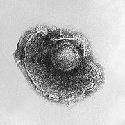
Herpes viruses have a lipid envelope
Helical
These viruses are composed of a single type of capsomer stacked around a central axis to form a helical structure, which may have a central cavity, or hollow tube. This arrangement results in rod-shaped or filamentous virions: these can be short and highly rigid, or long and very flexible. The genetic material, generally single-stranded RNA, but ssDNA in some cases, is bound into the protein helix by interactions between the negatively charged nucleic acid and positive charges on the protein. Overall, the length of a helical capsid is related to the length of the nucleic acid contained within it and the diameter is dependent on the size and arrangement of capsomers. The well-studied Tobacco mosaic virus is an example of a helical virus.
Icosahedral
Most animal viruses are icosahedral or near-spherical with icosahedral symmetry. A regular icosahedron is the optimum way of forming a closed shell from identical sub-units. The minimum number of identical capsomers required is twelve, each composed of five identical sub-units. Many viruses, such as rotavirus, have more than twelve capsomers and appear spherical but they retain this symmetry. Capsomers at the apices are surrounded by five other capsomers and are called pentons. Capsomers on the triangular faces are surround by six others and are call hexons.
Envelope
Some species of virus envelope themselves in a modified form of one of the cell membranes, either the outer membrane surrounding an infected host cell, or internal membranes such as nuclear membrane or endoplasmic reticulum, thus gaining an outer lipid bilayer known as a viral envelope. This membrane is studded with proteins coded for by the viral genome and host genome; the lipid membrane itself and any carbohydrates present originate entirely from the host. The influenza virus and HIV use this strategy. Most enveloped viruses are dependent on the envelope for their infectivity.
Complex
These viruses possess a capsid that is neither purely helical, nor purely icosahedral, and that may possess extra structures such as protein tails or a complex outer wall. Some bacteriophages, such as Enterobacteria phage T4 have a complex structure consisting of an icosahedral head bound to a helical tail, which may have a hexagonal base plate with protruding protein tail fibres. This tail structure acts like a molecular syringe, attaching to the bacterial host and then injecting the viral genome into the cell.

The poxviruses are large, complex viruses that have an unusual morphology. The viral genome is associated with proteins within a central disk structure known as a nucleoid. The nucleoid is surrounded by a membrane and two lateral bodies of unknown function. The virus has an outer envelope with a thick layer of protein studded over its surface. The whole virion is slightly pleiomorphic, ranging from ovoid to brick shape. Mimivirus is the largest known virus, with a capsid diameter of 400 nm. Protein filaments measuring 100 nm project from the surface. The capsid appears hexagonal under an electron microscope, therefore the capsid is probably icosahedral.

Some viruses that infect Archaea have complex structures that are unrelated to any other form of virus, with a wide variety of unusual shapes, ranging from spindle-shaped structures, to viruses that resemble hooked rods, teardrops or even bottles. Other archaeal viruses resemble the tailed bacteriophages, and can have multiple tail structures.

Genome

An enormous variety of genomic structures can be seen among viral species; as a group they contain more structural genomic diversity than plants, animals, archaea, or bacteria. There are millions of different types of viruses; though only about 5,000 of them have been described in detail. A virus has either DNA or RNA genes and is called a DNA virus or a RNA virus respectively. By far most viruses have RNA. Plant viruses tend to have single-stranded RNA and bacteriophages tend to have double-stranded DNA. Viral genomes are circular, such as polyomaviruses, or linear, such as adenoviruses. The type of nucleic acid is irrelevant to the shape of the genome. Among RNA viruses, the genome is often divided up into separate parts within the virion and is called segmented. Each segment often codes for one protein and they are usually found together in one capsid. Every segment is not required to be in the same virion for the overall virus to be infectious, as demonstrated by the brome mosaic virus. A viral genome, irrespective of nucleic acid type, is either single-stranded or double-stranded. Single-stranded genomes consist of an unpaired nucleic acid, analogous to one-half of a ladder split down the middle. Double-stranded genomes consist of two complementary paired nucleic acids, analogous to a ladder. Some viruses, such as those belonging to the Hepadnaviridae, contain a genome that is partially double-stranded and partially single-stranded. For viruses with RNA or single-stranded DNA, the strands are said to be either positive-sense (called the plus-strand) or negative-sense (called the minus-strand), depending on whether it is complementary to the viral messenger RNA (mRNA). Positive-sense viral RNA is identical to viral mRNA and thus can be immediately translated by the host cell. Negative-sense viral RNA is complementary to mRNA and thus must be converted to positive-sense RNA by an RNA polymerase before translation. DNA nomenclature is similar to RNA nomenclature, in that the coding strand for the viral mRNA is complementary to it (−), and the non-coding strand is a copy of it (+).Genome size varies greatly between species. The smallest viral genomes code for only four proteins and have a mass of about 106 Daltons; the largest have a mass of about 108 Daltons and code for over one hundred proteins. RNA viruses generally have smaller genome sizes than DNA viruses because of a higher error-rate when replicating, and have a maximum upper size limit. Beyond this limit, errors in the genome when replicating render the virus useless or uncompetitive. To compensate for this, RNA viruses often have segmented genomes where the genome is split into smaller molecules, thus reducing the chance of error. In contrast, DNA viruses generally have larger genomes because of the high fidelity of their replication enzymes.
A cartoon showing how viral genes can be shuffled to form new viruses
How antigenic shift, or reassortment, can result in novel and highly pathogenic strains of human influenza
Viruses undergo genetic change by several mechanisms. These include a process called genetic drift where individual bases in the DNA or RNA mutate to other bases. Most of these point mutations are "silent"—they do not change the protein that the gene encodes—but others can confer evolutionary advantages such as resistance to antiviral drugs.Antigenic shift occurs when there is a major change in the genome of the virus. This can be a result of recombination or reassortment. When this happens with influenza viruses, pandemics might result. RNA viruses often exist as quasispecies or swarms of viruses of the same species but with slightly different genome nucleoside sequences. Such quasispecies are a prime target for natural selection.Segmented genomes confer evolutionary advantages; different strains of a virus with a segmented genome can shuffle and combine genes and produce progeny viruses or (offspring) that have unique characteristics. This is called reassortment or viral sex Genetic recombination is the process by which a strand of DNA is broken and then joined to the end of a different DNA molecule. This can occur when viruses infect cells simultaneously and studies of viral evolution have shown that recombination has been rampant in the species studied. Recombination is common to both RNA and DNA viruses.

Replication cycle

Viral populations do not grow through cell division, because they are acellular; instead, they use the machinery and metabolism of a host cell to produce multiple copies of themselves, and they assemble in the cell.

A typical virus replication cycle
Some bacteriophages inject their genomes into bacterial cells

The life cycle of viruses differs greatly between species but there are six basic stages in the life cycle of viruses:

  • Attachment is a specific binding between viral capsid proteins and specific receptors on the host cellular surface. This specificity determines the host range of a virus. For example, HIV infects only human T cells, because its surface protein, gp120, can interact with CD4 and receptors on the T cell's surface. This mechanism has evolved to favour those viruses that only infect cells in which they are capable of replication. Attachment to the receptor can induce the viral-envelope protein to undergo changes that results in the fusion of viral and cellular membranes.
  • Penetration follows attachment; viruses enter the host cell through receptor mediated endocytosis or membrane fusion. This is often called viral entry. The infection of plant cells is different from that of animal cells. Plants have a rigid cell wall made of cellulose and viruses can only get inside the cells after trauma to the cell wall. Viruses such as tobacco mosaic virus can also move directly in plants, from cell to cell, through pores called plasmodesmata. Bacteria, like plants, have strong cell walls that a virus must breach to infect the cell. Some viruses have evolved mechanisms that inject their genome into the bacterial cell while the viral capsid remains outside.
  • Uncoating is a process in which the viral capsid is degraded by viral enzymes or host enzymes thus releasing the viral genomic nucleic acid.
  • Replication involves synthesis of viral messenger RNA (mRNA) for viruses except positive sense RNA viruses (see above), viral protein synthesis and assembly of viral proteins and viral genome replication.
  • Following the assembly of the virus particles, post-translational modification of the viral proteins often occurs. In viruses such as HIV, this modification (sometimes called maturation) occurs after the virus has been released from the host cell.
  • Viruses are released from the host cell by lysis—a process that kills the cell by bursting its membrane. Enveloped viruses (e.g., HIV) typically are released from the host cell by budding. During this process the virus acquires its envelope, which is a modified piece of the host's plasma membrane.

The genetic material within viruses, and the method by which the material is replicated, vary between different types of viruses.

DNA viruses
The genome replication of most DNA viruses takes place in the cell's nucleus. If the cell has the appropriate receptor on its surface, these viruses enter the cell by fusion with the cell membrane or by endocytosis. Most DNA viruses are entirely dependent on the host cell's DNA and RNA synthesising machinery, and RNA processing machinery. The viral genome must cross the cell's nuclear membrane to access this machinery.
RNA viruses
These viruses are unique because their genetic information is encoded in RNA. Replication usually takes place in the cytoplasm. RNA viruses can be placed into about four different groups depending on their modes of replication. The polarity (whether or not it can be used directly to make proteins) of the RNA largely determines the replicative mechanism, and whether the genetic material is single-stranded or double-stranded. RNA viruses use their own RNA replicase enzymes to create copies of their genomes.
Reverse transcribing viruses
These replicate using reverse transcription, which is the formation of DNA from an RNA template. Reverse transcribing viruses containing RNA genomes use a DNA intermediate to replicate, whereas those containing DNA genomes use an RNA intermediate during genome replication. Both types use the reverse transcriptase enzyme to carry out the nucleic acid conversion. Retroviruses often integrate the DNA produced by reverse transcription into the host genome. They are susceptible to antiviral drugs that inhibit the reverse transcriptase enzyme, e.g. zidovudine and lamivudine. An example of the first type is HIV, which is a retrovirus. Examples of the second type are the Hepadnaviridae, which includes Hepatitis B virus.

Effects on the host cell

The range of structural and biochemical effects that viruses have on the host cell is extensive. These are called cytopathic effects. Most virus infections eventually result in the death of the host cell. The causes of death include cell lysis, alterations to the cell's surface membrane and apoptosis. Often cell death is caused by cessation of its normal activities because of suppression by virus-specific proteins, not all of which are components of the virus particle.

Some viruses cause no apparent changes to the infected cell. Cells in which the virus is latent and inactive show few signs of infection and often function normally. This causes persistent infections and the virus is often dormant for many months or years. This is often the case with herpes viruses. Some viruses, such as Epstein-Barr virus, can cause cells to proliferate without causing malignancy, while others, such as papillomaviruses, are established causes of cancer.